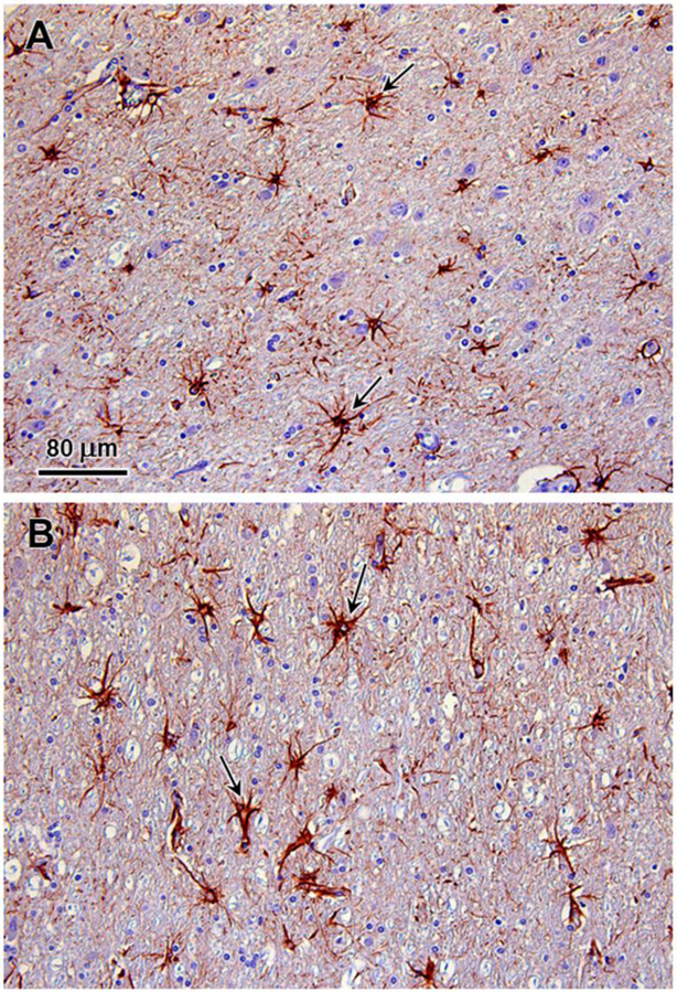
Fig. 4.
Immunohistochemical demonstration of reactive astrocytes in the parietal cerbral cortex of the affected dog. GFAP immunostaining of sections of the cerebral cortex demonstrated numerous reactive astrocytes (arrows) in cortical layers 6A (polymorphic cell layer) (A) and 6B (B). Reactive astrocytes were sparse elsewhere in the cerebral cortex.

