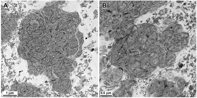
Fig. 9.
Electron micrographs of storage bodies in cerebellar Purkinje cells of the affected dogaffected dog. As with the cerebral cortical neurons, the contents of most storage bodies consisted of tightly packed clusters of membrane-like structures, but none of the electron dense amorphous patches of material were observed in the Purkinje cell storage bodies. As with the cerebral cortex, membranes enclosing the storage material (arrows) were only partially preserved due to the initial fixation in formalin.

