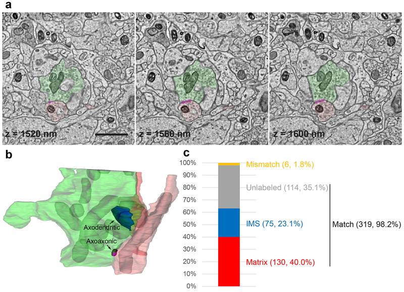
Figure 4. Multiplexed peroxidase labeling in volume EM.
(a) Three consecutive sections from one of the samples shown in Fig. 3b in which spinal cord dorsal horn inhibitory interneurons (mitochondrial matrix) were labeled using Slc32a1IRES-Cre and AAV1-DIO-Matrix-dAPEX2 (axon in light red and dendrite in dark red), and primary somatosensory afferents (mitochondrial IMS) were labeled using AvilFlpO and AAV9-FDIO-IMS-dAPEX2 (green). Magenta overlay: axoaxonic synapse between an inhibitory interneuron and the primary afferent. The z coordinates from the top of the volume are noted on each image. Scale bar: 1 μm. See also Supplementary Video 1.
(b) The 3D reconstruction of the same primary afferent and inhibitory interneuron profiles. Labeled mitochondria (grey) and an axodendritic synapse between the primary afferent and an inhibitory interneuron (blue) are additionally reconstructed. See also Supplementary Video 2.
(c) Level of concordance between independent annotations of mitochondria (matrix-labeled, IMS-labeled, or unlabeled) in a volume of 12 × 8 × 2 μm by two annotators. The numbers of each category as well as their proportions of the total number of mitochondria are indicated in parentheses. The three categories Matrix, IMS, and Unlabeled all contain matching annotations, while the Mismatch category contains mismatching annotations.

