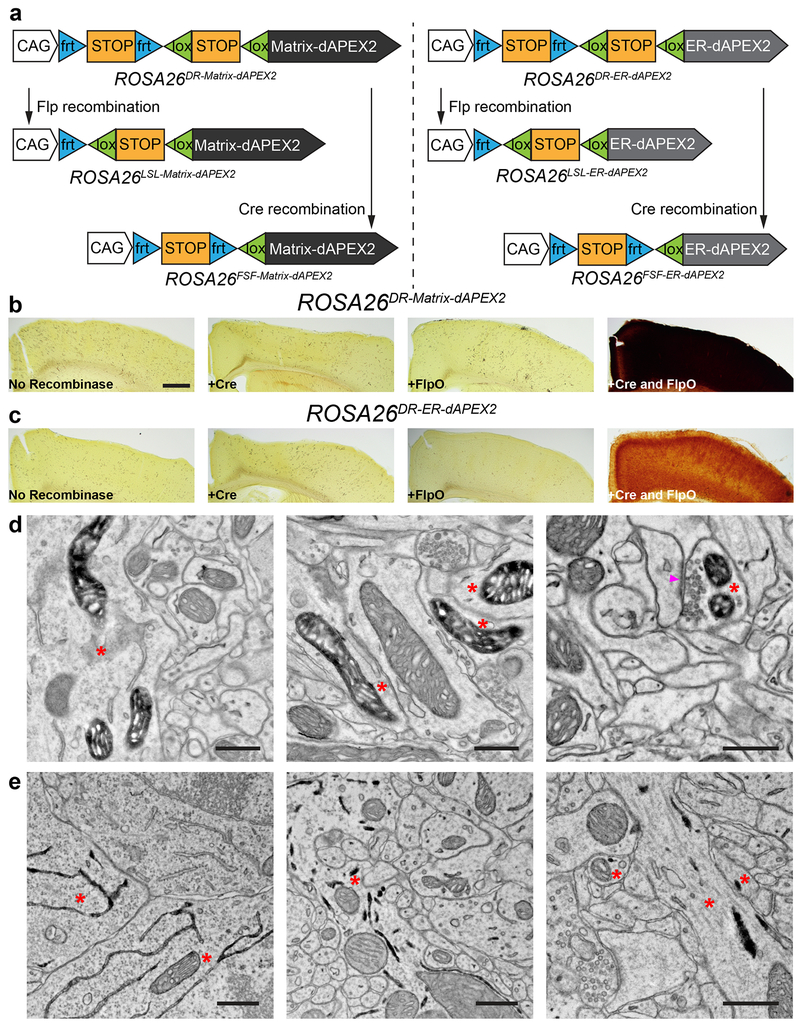
Figure 5. Generation of recombinase-dependent mouse dAPEX2 reporter lines.
(a) Schematics showing overviews of the six mouse reporter lines. Single-recombinase-dependent lines were generated by germline deletion of one of the STOP cassettes.
(b, c) LM images showing cortical sections after injections of AAVs encoding various recombinases into dual-recombinase-dependent ROSA26DR-Matrix-dAPEX2 (b) and ROSA26DR-ER-dAPEX2 animals (c). Only endogenous peroxidase activity was observed when no recombinase, Cre alone, or FlpO alone was transduced (left three panels). dAPEX2 peroxidase staining was observed only following co-injection of Cre and FlpO viruses (rightmost panels).
(d) EM images from the cortex of a ROSA26DR-Matrix-dAPEX2 animal co-transduced with Cre and FlpO. Asterisks: labeled neurons. Labeled mitochondria can be seen in soma, dendrites, and axons, consistent with results using AAVs to express peroxidase constructs. Arrowhead: synapse made by the labeled neuron. n = 4 animals and experiments.
(e) EM images of the cortex of a ROSA26DR-ER-dAPEX2 animal co-transduced with Cre and FlpO. Asterisks: labeled neurons. Labeled ER can be seen in somata and dendrites, as expected. n = 4 animals and experiments.
Scale bars: b, c: 500 μm, d, e: 0.5 μm.

