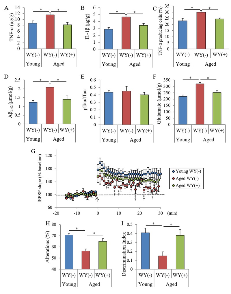
Figure 4.

Effects of WY peptides on cognitive decline in aged mice. Seven and 68-week-old male C57BL/6J mice were maintained on a diet with or without 0.05% (w/w) WY peptide for 19 weeks. (A, B) The levels of TNF-α and IL-1β in the hippocampus. (C) Characterization of CD11b-positive microglia in the brain isolated by MACS and flow cytometry. The percentages of TNF-α-producing cells to CD11b-positive cells. (D-F) The levels of TBS-soluble Aβ1-42, pTau (pS199)/total Tau, and glutamate in the hippocampus. (G) LTP measurement from a hippocampus slice after theta-burst stimulation (arrow). (H) Spontaneous alterations during 8 min of exploring in Y-maze. (I) Discrimination index in NORT by measuring the time spent exploring novel and familiar objects during 5 min of re-exploration. Data are the mean ± SEM of 7-week-old mice (n = 15) fed with control diet, 68-week-old mice (n = 13) fed with control diet, and 68-week-old mice (n = 10) fed with diet containing WY peptide. p-values shown in the graph were calculated by one-way ANOVA followed by the Tukey–-Kramer test. p-values shown in the graph were calculated by unpaired t-test in (G). *p < 0.05,†p < 0.05.
