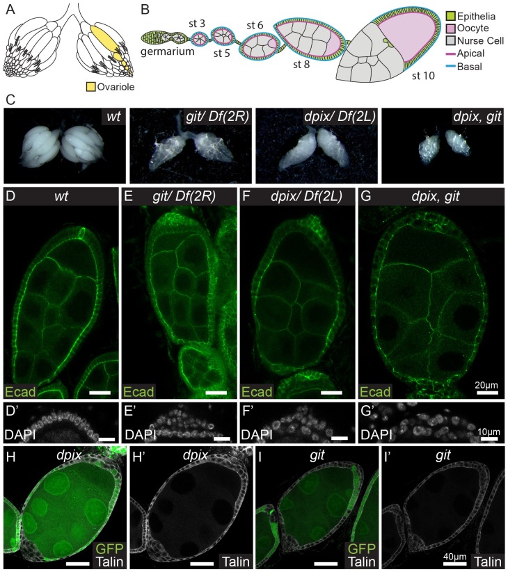
Fig 1. The dPix-Git complex is required cell autonomously for epithelial morphogenesis during Drosophila melanogaster egg chamber development.
(A-B) Schematic diagrams of an adult D. melanogaster ovary with an ovariole structure highlighted (yellow) (A), and an individual ovariole containing egg chambers up to stage 10 (B). Key tissue types are follicular epithelia (green), oocyte (pink), and germline nurse cells (grey). (C) General appearance of adult D. melanogaster ovaries of the indicated genotypes: from left to right, wild-type (wt); git; dpix; dpix, git. (D-G’) Stage 7–8 egg chambers stained with E-Cadherin (Ecad), and enlarged projections of the posterior tip of egg chambers stained with DAPI (D’, E’, F’, G’) for the following genotypes: (D-D’) wild-type; (E-E’) git; (F-F’) dpix; (G-G’) dpix, git. Scale bars 20 μm for (D, E, F, G) and 10 μm for (D’, E’, F’, G’). (H-I’) D. melanogaster egg chambers with a mosaic of wild-type and dpix (H-H’) or git (I-I’) mutant tissue. In each egg chamber wild-type tissue is labelled by GFP, whereas dpix and git mutant tissue lacks GFP. Cells are visualised with Talin antibody (grey). Scale bars 40 μm.

