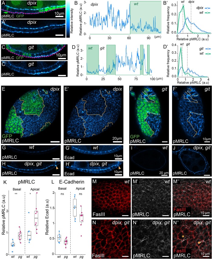
Fig 3. The dPix-Git complex spatially restricts subcellular activation of myosin to maintain tissue integrity during epithelial development.
(A-F’) Comparison of phosphorylated myosin regulatory light chain (pMRLC), between wild-type and adjacent mutant tissue. For (A, C, E, F) mutant tissue lacks GFP. (A-A’) Cross-section of a stage 6–7 egg chamber, with a mosaic of wild-type and dpix tissue. (B-B’) Quantification of pMRLC intensity (B) along a basal transect (magenta line in A) of dpix mosaic tissue from (A-A’), and corresponding normalised frequency polygons depicting distribution of pMRLC values in each genotype (B’). Dashed lines in (B’) are average intensity values for the indicated genotype. Bin width = 0.2. (C-C’) Basal cross-section of a stage 6 egg chamber, with a mosaic of wild-type and git tissue. (D-D’) Quantification of pMRLC intensity (D) along a basal transect (magenta line in C) of git mosaic tissue from (C-C’), and corresponding normalised frequency polygons depicting distribution of pMRLC values in each genotype (D’). Dashed lines in (D’) are average intensity values for the indicated genotype. Bin width = 0.2. (E-E’) Basal pMRLC distribution in a dpix mosaic tissue that has maintained monolayering in main body follicle cells. (F-F’) Basal pMRLC distribution in git mosaic tissue with multilayering in git mutant tissue. Orange lines in (E-F’) indicate genotype boundaries. (G-J) Follicular epithelia of stage 6–7 wild-type and dpix, git double mutant egg chambers co-stained with pMRLC and E-Cadherin (Ecad). (G-G’) wild-type egg chamber. (H-H’) dpix, git mutant egg chamber. Arrowhead indicates collapsed lateral membrane at the point of pMRLC accumulation in dpix, git mutant. (I) Tissue scale cross section of pMRLC signal from wild-type egg chamber in (G-G’). (J) Tissue scale cross section of pMRLC signal from dpix, git egg chamber in (H-H’). Scale bars 10 μm (G-H’), and 20 μm (I-J). (K-L) Quantification of apical and basal pMRLC, and E-Cadherin in wild-type and dpix, git double mutant follicular epithelia. Genotypes and sample sizes are: wild-type (wt), n = 6 egg chambers; dpix, git (pg), n = 8 egg chambers. Statistical analyses are Welch’s unequal variances t-tests. Significance: * = p < 0.05; ** = p < 0.01; ns = not significant. (M-N”) Basal sections of ~stage 7 wild-type (M-M”) and dpix, git (N-N”) egg chambers. Membranes are marked by fasciclin III (FasIII) and activated myosin is marked by pMRLC. Purse-string like accumulation of pMRLC is indicated by asterisk, and aberrant-cell junction indicated by arrowhead (N”). Relative intensity index applies to pMRLC and Ecad signal in (A-A’, C-C’, E-J).

