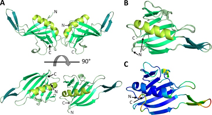
Fig 1. Atomic structure of RVFV L protein C-terminal domain.
A) The structure of the protein dimer in the asymmetric unit is shown as a ribbon diagram in front and top view. Corresponding structural elements are shown in the same color (large β-sheet in medium green, long α-helix in lime green, β-hairpin in teal). N- and C-termini are labelled. B) Structures of RVFV CBD chain A and chain B are superimposed and depicted with the same color code. C) Representation of chain A of RVFV CBD as a ribbon diagram colored by B-factor with the highest observed B being 26 (orange) and the lowest 1 (dark blue).

