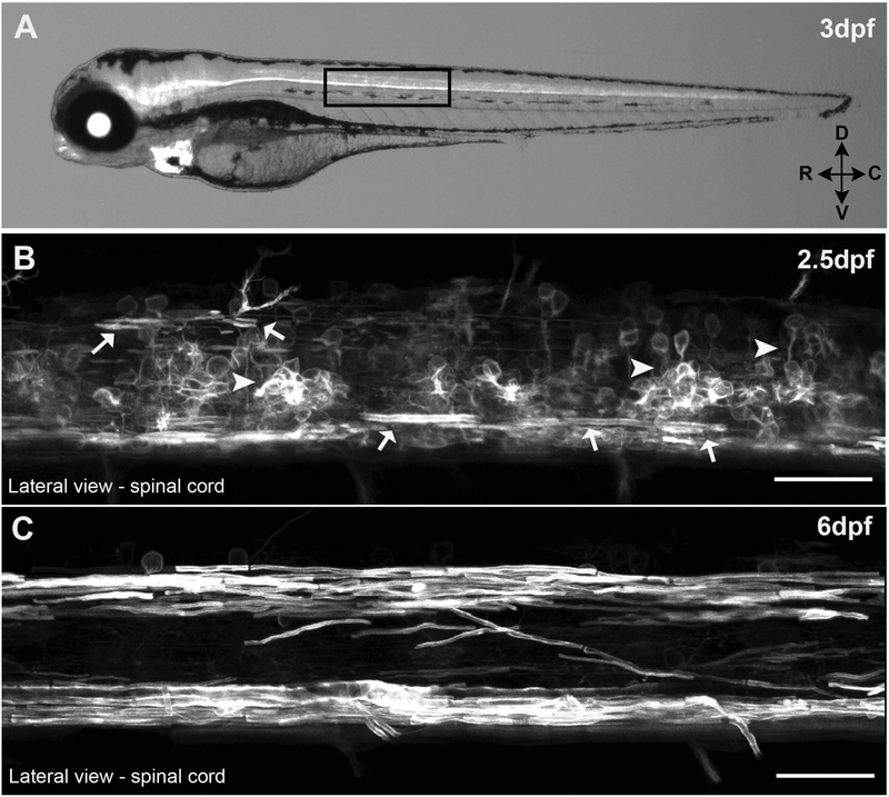
Figure 1: Tg(mpz:mEGFP) fish.
(A) 3 day post fertilization (dpf) larvae expressing membrane tethered EGFP under the control of a 12.5kb mpz promoter fragment. High magnification confocal z-stack images of the lateral spinal cord show some myelinating oligodendrocytes (arrows) at 2.5dpf (B) and robustly myelinated spinal tracks by 6 dpf (C) in live larvae. (scale bar = 25μM)

