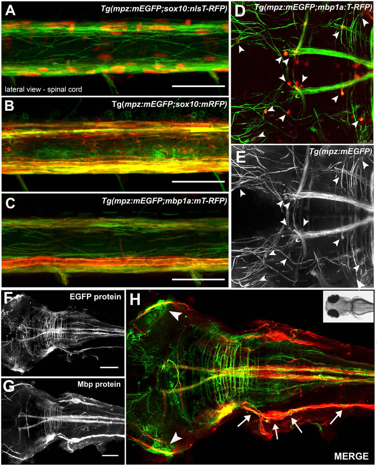
Figure 2: The mpz:mEGFP reporter co-localized with known oligodendrocyte markers.
mpz reporter expression (green) co-labeled oligodendrocyte lineage cells in the spinal cord at 6dpf in Tg(sox10:T-RFPnls+) (red nuclei, A), Tg(sox10:mRFP+) (red membranes, B) and Tg(mbp1a:mRFP) (red membranes, C) transgenic lines, imaged laterally by live confocal microscopy. Similarly, T-RFP positive cell bodies (arrowheads, D) also were faintly mEGFP positive (arrowheads D, E) within mEGFP-positive myelinated tracks in the brain of a 6dpf Tg(mpz:mEGFP;mbp1a:T-RFP)casper larvae, imaged dorsally by live confocal microscopy. Whole mount immmunohistochemistry for EGFP protein (green F,H) and Mbp protein (G,H red) was consistent with mpz reporter expression specifically in myelinating glia in the brain, spinal cord, lateral lines (arrows) and cranial nerves (arrowheads) of 6dpf Tg(mpz:mEGFP) larvae, although MBP expression was much stronger in lateral line than mpz reporter expression, consistent with low expression of mpz in Schwann cells at this stage. (scale bar 50μM, A-C; 100μM, D-F, 25μM, A-C).

