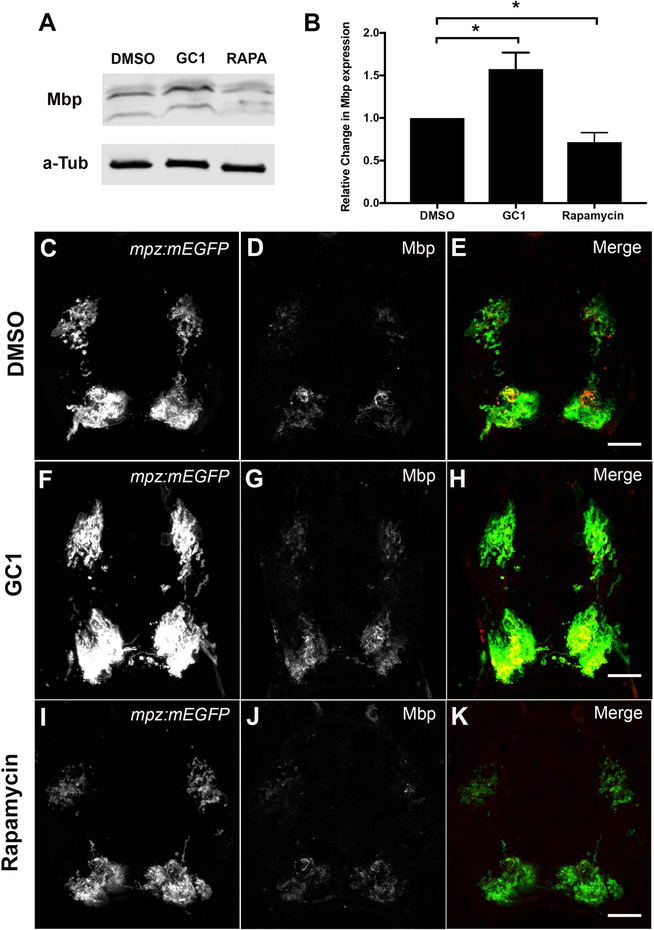
Figure 5: Western blot analysis and immunohistochemistry for Mbp protein expression correlated with mpz reporter activity in drug-treated larvae.
(A) Representative western blot of Mbp protein expression in 2–4dpf DMSO, GC1 (10nM), or rapamycin (20μM) treated larvae. (B) Quantification of western blots showed that GC1 treatment significantly increased Mbp expression relative to DMSO treated controls (alpha tubulin loading control), while rapamycin significantly decreased Mbp protein expression in developing larvae. Immunohistochemistry of transverse spinal cord sections of 6dpf Tg(mpz:mEGFP) larvae treated with DMSO (C-E), GC1 (F-H) or rapamycin (I-K) showed changes in Mmb and EGFP expression in myelinated tracts in response to drug treatments. (Scale bar = 10μM; statistical analysis: 1-way Anova, p<0.05* <0.01** <0.001*** <0.0001****)

