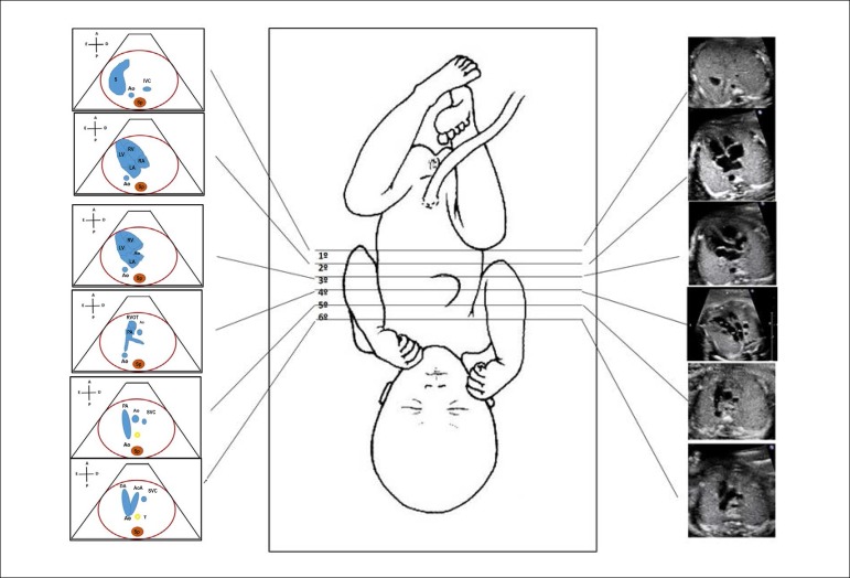
Figure 2.1.
Standardization of fetal heart screening, scanning the fetal vessels and heart from the infradiaphragmatic region towards the cranium. There are 6 levels, being the first exactly below the diaphragm, which allows the identification of the descending aorta and inferior vena cava; second, the four-chamber view; third, left ventricular outflow tract; fourth, right ventricular outflow tract; fifth, three vessel view, and, sixth, three vessel and trachea view.
Ao: Aorta; AoA: aortic arch; Asc: ascending; DA: ductus arteriosus; IVC: inferior vena cava; LA: left atrium; LV: left ventricle; PA: pulmonary artery; RA: right atrium; RV: right ventricle; RVOT: right ventricular outflow tract; S: stomach; Sp: spine; SVC: superior vena cava; T: trachea.

