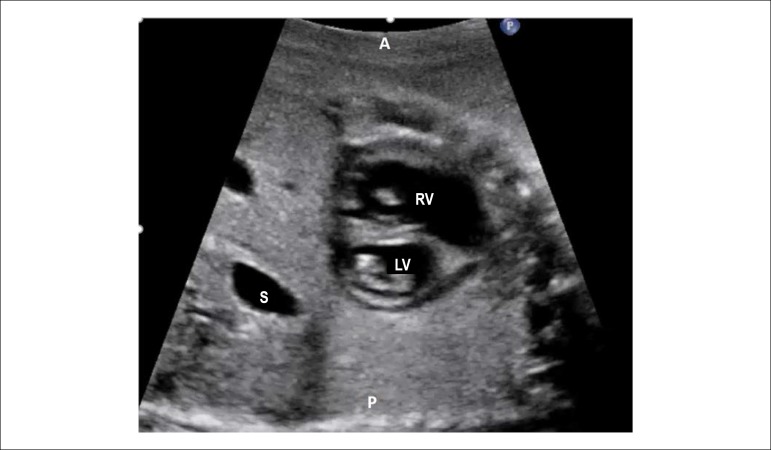
Figure 2.6.
Short-axis of the ventricles. In this plane it is possible to analyze the position of the papillary muscles of the right and left ventricles. It is also of great utility in detecting subtler forms of atrioventricular septal defect when it is presented with two valvular orifices.
A: anterior; P: posterior; LV: left ventricle; RV: right ventricle; S: stomach.

