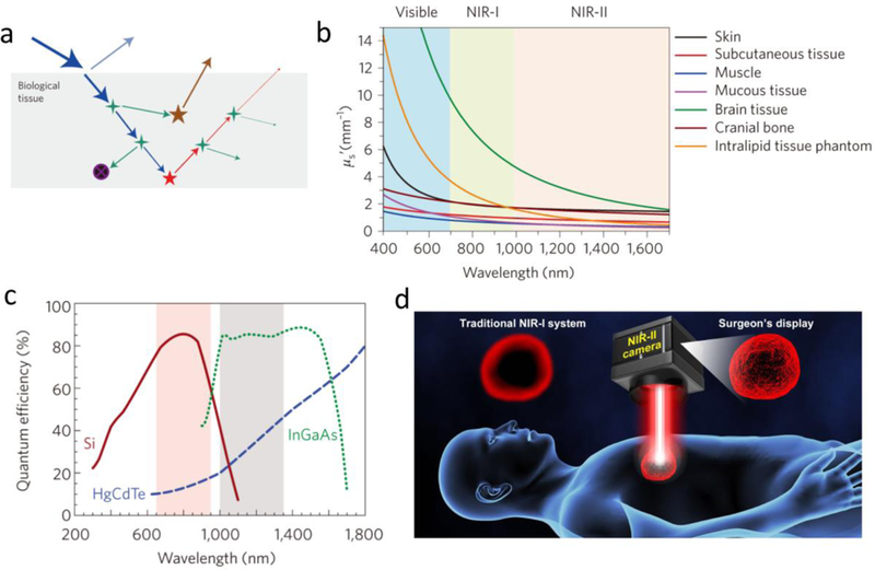
Figure 15. The reduced tissue autofluorescence and scattering are the motivation for developing NIR-II fluorescence-derived biomedical imaging.

a) Excitation laser-organ/tissue interactions include blue excitation light, cyan reflection, green scattering, black absorption, as well as brown autofluorescence. All of these parameters generate the loss of fluorescence and the gain of background signals (noise). b) Diminished scattering coefficients of different tissue phantoms as a function of wavelengths. c) Quantum efficiency curves for several cameras based on silicon, InGaAs or HgCdTe sensors. d) NIR-II guided imaging will improve the surgery accuracy with lower autofluorescence and scattering compared with NIR-I navigation system. Reproduced with permission from ref. [4] for a-b) and ref. [150] for c).
