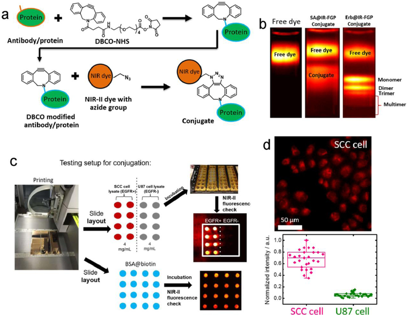
Figure 7. Bioconjugations between NIR-II fluorophores and proteins.

a) The copper-free click chemistry was applied to conjugate the IR-FGP fluorophore with proteins of interest. b) Density gradient ultracentrifugation (DGU) was performed to purify the SA@IR-FGP and Erb@IR-FGP conjugates. The applied sucrose gradient encompasses from 1.06–1.23 g/ cm3 (15–50 wt.%) (imaging details: 850/1000 nm short-pass (SP) filters and 900/1100-nm LP emission filters). c) A new reverse phase protein lysate microarray method was exploited to test the quality of conjugates. In detail, 1. The BSA-biotin, “catching” a primary antibody or cell lysate, was printed on the gold chips. 2. The purified conjugates were incubated on top of the printed spots under the cavity of frames. 3. Check the PL intensity by 10X magnification NIR II set-up with desired long-pass emission filter combination. d) Targeted cell imaging by Erb@IR-FGP on positive and negative cell lines respectively (imaging details: 785 nm excitation and 1050 nm LP emission filter). Reprinted with permission from Ref. [93].
