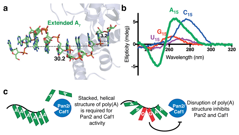
Figure 7. Model for the recognition of the intrinsic poly(A) structure by Pan2 and Caf1 deadenylases.
a, Extended oligo(A) helix bound to Pan2, modeled by duplicating and superposing the observed A5, showing the helical conformation adopted by oligo(A) in the active site. Distances are shown in Ångstroms. b, Circular dichroism spectra of A15 (green), U15 (purple), C15 (blue), and G15 (red) RNAs. These spectra are representative of identical experiments performed 2 times. c, Proposed model for the recognition of poly(A) RNA by the Caf1 and Pan2 DEDD family deadenylases.

