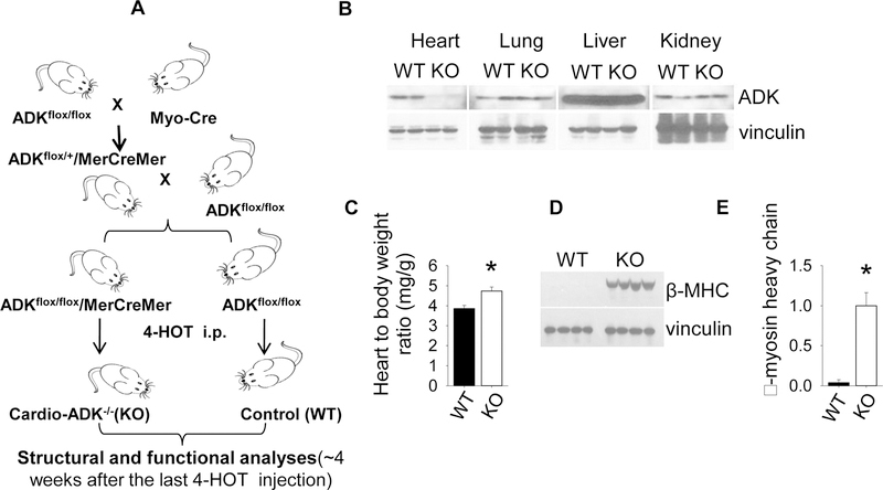
Figure 1.
Cardiomyocyte specific ADK disruption causes spontaneous LV hypertrophy. (A) Tamoxifen induced cardiomyocyte specific excision of floxed exon 7 in the ADK gene. (B) 4 weeks after the last tamoxifen injection, ADK protein expression was eliminated specifically in heart muscle in cADK−/− mice. (C) Heart weight to body weight ratio of cADK−/− and WT mice (n=9 WT and 10 cADK−/− ). (D, E) β-MHC expression in WT and cADK−/− was analyzed by western blot (n=4 WT and 4 ADK−/−).

