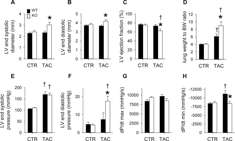
Figure 3. Effects of cardiomyocyte ADK disruption on LV function during pressure overload.
(A) Echocardiography measurements of end systolic diameter (ESD), (B) end diastolic diameter (EDD) and (C) ejection fraction (EF) 6 weeks after sham or TAC surgery. (n=10, 11, 12, and 14 for WT. ADK−/−, WT-TAC, and ADK−/−-TAC, respectively) (D) Lung weight to body weight ratio in WT and cADK−/− mice 6 weeks after TAC (n= 26, 24, 17, and 17 for, WT, ADK−/−, WT-TAC, and ADK−/−-TAC, respectively). (E) Left ventricular end systolic and (F) diastolic pressures and rates of LV pressure development during (G) systole and (H) diastole in WT and cADK−/− mice after TAC. (n=8, 9, 6, and 10 for WT. ADK−/−, WT-TAC, and ADK−/−-TAC, respectively)

