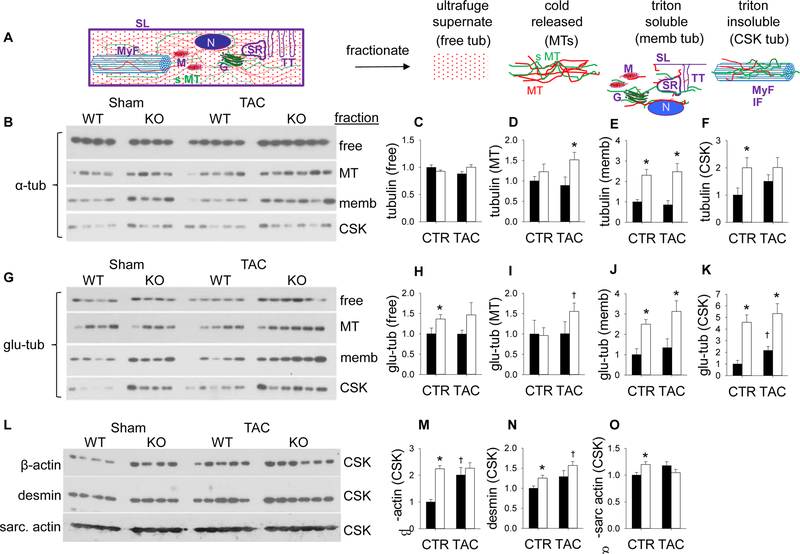
Figure 5. ADK disruption increases microtubule stabilization/detyrosination.
(A) Ventricular lysates were separated into different Free, Microtubule (MT), Membrane (Memb), and Cytoskeletal (CSK) fractions as depicted. (N is nucleus, G is golgi, SR is sarcoplasmic reticulum, TT is t-tubules, M is mitochondria, SL is sarcolemma, CSK is cytoskeleton, MT is microtubule, and sMT is stabilized microtubule). Cardiac lysates were analyzed by western blot for alpha tubulin (B–F) and detyrosinated tubulin (Glu-tubulin) (G–K). β-actin (L,M), desmin (L, N) and sarcomeric actin (L,O), were also measured in the triton insoluble cytoskeletal fraction. (n=4, 4, 5, and 6 for WT. cADK−/−, WT-TAC, and cADK−/− TAC respectively).

