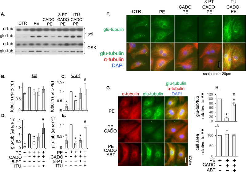
Figure 6. Pharmacological inhibition of ADK reverses CADO attenuation of microtubule stabilization/detyrosination.
(A) Neonatal cardiomyocytes (NRVMs) were treated with phenylephrine (50 μM) for 48 hours in presence of CADO (5 μM), CADO + non-selective adenosine receptor antagonist 8-PT (10 μM), or CADO + ADK inhibitor iodotubercidin (ITU; 0.3 µM). α-tubulin (B and C) and detyrosinated α-tubulin (glu-tubulin) (D and E) were measured by western blot in triton soluble (sol) (B and D) and insoluble (CSK) (C and E) fractions (n ≥ 4 per condition. * indicates p < .05 compared to PE. # indicates p < .05 compared to PE-CADO ). (F) Immunofluorescence staining of NRVMs for α-tubulin and glu-tubulin after treatment described above. (G) NRVMs were treated with PE for 48 hours to induce hypertrophy, followed by an additional 24 hours with PE, PE + CADO, or PE + CADO + ABT-702(0.3 μM). α-tubulin and glu-tubulin were visualized by immunofluorescence and the area of detyrosinated tubulin was divided by the area of total tub (H) in 25–30 cells per condition. Percent change in cell area was also measured (J). Results are the average of 3 experiments, relative to continued PE treatment alone

