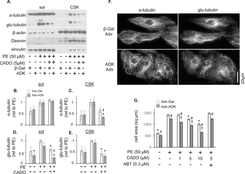
Figure 7. ADK adenovirus augments CADO suppression of microtubule stabilization/detyrosination.
(A) Neonatal cardiomyocytes (NRVMs) were infected with β-gal or ADK expressing adenovirus (adv) for 24 hours prior to treatment with PE for 48 hours in the presence or absence of CADO (5 μM). Soluble (A, B, D) and cytoskeletal fractions (B, C, E) were examined by western blot for α-tubulin and glu-tubulin. CADO + non-selective adenosine receptor antagonist 8-PT (10 μM), or CADO + ADK inhibitor iodotubercidin (ITU; 0.3 μM). α-tubulin (B and C) and detyrosinated α-tubulin (glu-tubulin) (D and E) were measured by western blot in triton soluble (sol) (B and D) and insoluble (CSK) (C and E) fractions (n ≥ 4 per condition. * indicates p<.05 compared to PE. † indicates p<.05 compared to PE-CADO). (F) NRVMs infected with β-gal or ADK adv were examined by immunofluorescence for α-tubulin and glu-tubulin. (G) Cell area was measured in NRVMs infected with β-gal or ADK adv 48 hours after treatment with PE, PE + 1, 5, or 10 M CADO, or PE + 5 μM CADO + 0.3 μM ABT-702. (Bars represent the average cell area of at least 100 cells measured per condition; (* indicates p<.05 compared to PE. indicates p<.05 compared to PE-CADO. # indicates p<.05 comparing ADK adv to β-gal adv under same treatment)

