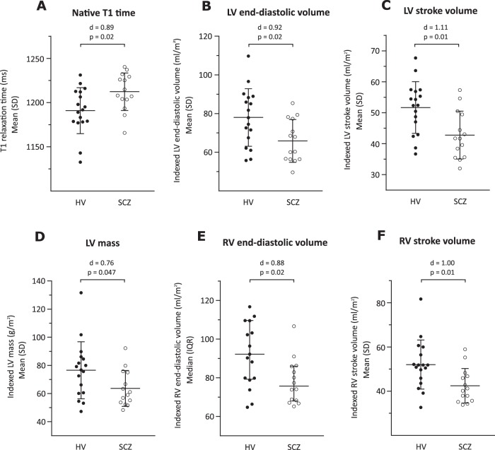
Fig. 3. Cardiac structure and function in patients with schizophrenia (SCZ) and healthy volunteers (HV).
a Native myocardial T1 time was longer in SCZ compared with HV (d = 0.89; p = 0.02); b Indexed left ventricular (LV) end-diastolic volume was smaller in SCZ compared with HV (d = 0.92; p = 0.02); c Indexed LV stroke volume was smaller in SCZ compared with HV (d = 1.11; p = 0.01); d Indexed LV mass was lower in SCZ compared with HV (d = 0.76; p = 0.047); e Indexed right ventricular (RV) end-diastolic volume was smaller in SCZ compared with HV (d = 0.88; p = 0.02); f Indexed RV stroke volume was smaller in SCZ compared with HV (d = 1.00; p = 0.01). These findings are consistent with early diffuse myocardial fibrosis in patients with schizophrenia. d = Cohen’s d effect size for group mean difference. P values correspond to the unpaired t-test or Mann Whitney U test used to compare the 2 groups

