
Figure 4.
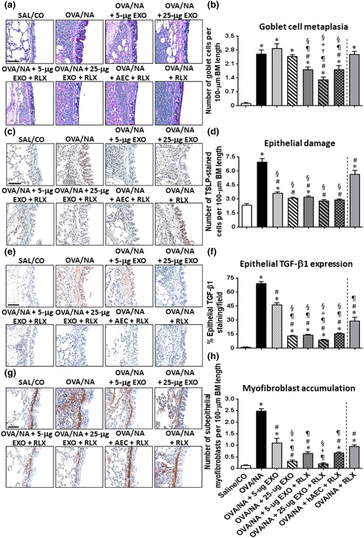
The effects of exosomes (EXO) on chronic AAD‐induced airway remodelling in the absence or presence of serelaxin (RLX). Representative images of ABPAS‐stained lung sections from mice subjected to OVA/NA‐induced chronic AAD and the various treatment investigated (a) demonstrate the extent of goblet cell metaplasia within the airways. Scale bar = 100 μm. Representative images of immunohistochemically stained lung sections from mice subjected to OVA/NA‐induced chronic AAD and the various treatment investigated show the extent of TSLP‐associated epithelial damage (c); epithelial TGF‐β1 expression (e); and subepithelial myofibroblast accumulation (g). Scale bar = 50 μm (c, e, g). In each case, the effects of serelaxin alone (previously evaluated in this model over the same time period; Patel et al., 2016) have been included to provide comparisons to the combination treatment groups evaluated in the current study. Also shown is the mean ± SEM number of goblet cells per 100‐μm BM length, as mean ± SEM, induced by OVA/NA (b); number of TSLP‐stained cells per 100‐μm BM length (d); % epithelial TGF‐β1 expression levels per field (f); and number of subepithelial myofibroblasts per 100‐μm BM length (h) from five airways per mouse; n = 8 animals per group. *P < 0.05, significantly different from the control group (SAL/CO); # P < 0.05, significantly different from the OVA/NA group; ¶ P < 0.05, significantly different from the OVA/NA + 5‐μg EXO‐treated group; † P < 0.05, significantly different from the OVA/NA + 25‐μg EXO‐treated group; + P < 0.05, significantly different from the OVA/NA + AEC + RLX‐treated group; § P < 0.05, significantly different from the OVA/NA + RLX‐treated group