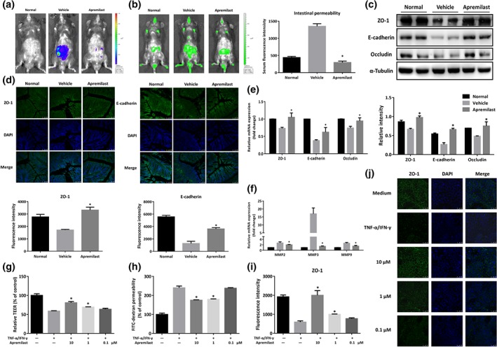
Figure 3.

Apremilast protected the intestinal epithelial barrier function and prevented cytokine‐induced epithelial barrier disruption. (a) Bioluminescent imaging with L‐012 sodium was obtained under isoflurane anaesthesia using an IVIS Spectrum CT system. (b) Fluorescence imaging with FITC‐dextran administration (left) and serum fluorescence intensity of FITC‐dextran (right) were measured. (c) The expression of tight junction‐associated proteins (ZO‐1, E‐cadherin, and occludin) detected by western blot and α‐tubulin was used as a loading control. (d) Colonic tissues were immunofluorescently stained for ZO‐1 and E‐cadherin, and the nuclei were stained with DAPI. (e) The mRNA level of tight junction‐associated proteins. (f) The mRNA expression of MMP2, MMP3, and MMP9. (g) Barrier function was measured as TEER in Caco‐2 cell monolayers primed by TNF‐α and IFN‐γ. (h) FITC‐dextran permeability in cytokine‐induced Caco‐2 cells. (i) Quantification of fluorescence intensity of ZO‐1 in Caco‐2 cells. (j) Representative image of immunofluorescent staining for ZO‐1 in Caco‐2 cells. Data shown are means ± SEM. (a–f), n = 8 per group. *P < 0.05, significantly different from vehicle (DSS only) group. (g–j), n = 5. *P < 0.05, significantly different from TNF‐α plus IFN‐γ‐treated group
