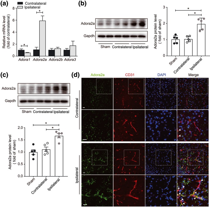
Figure 1.

Increased expression of A2A receptors in ischaemic brain. (a) Real‐time PCR analysis of Adora1, Adora2a, Adora2b, and Adora3 mRNA expression in whole brain 24 hr after eMCAo in mice (n = 6 mice per group). (b) Western blot analysis and densitometric quantification of A2A receptor protein expression in whole brain 24 hr after eMCAo (n = 5 mice per group). (c) Western blot analysis and densitometric quantification of A2A receptor protein expression in isolated BMECs, 24 hr after eMCAo (n = 5 per group). (d) Representative images of A2A receptors in the contralateral and ipsilateral cerebral hemispheres in mice with eMCAo. Brain sections were stained for A2A receptors (green), CD31 (red, microvessel), and DAPI (blue, nuclei). Images in the second and fourth rows are magnification of the boxed regions of images in the first and third rows respectively. Scale bar: 20 μm. Data are represented as means ± SD (for a, b) and ± SEM (for c). *P < .05, significantly different as indicated; unpaired Student's t test (for a) and one‐way ANOVA followed by Bonferroni's test (for b, c)
