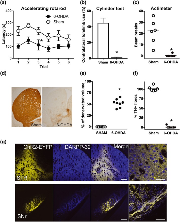
Figure 2.

Efficacy of dopamine lesions and channelrhodopsin viral infection. (a–c) 6‐OHDA‐lesioned animals showed motor coordination deficits (a; *P < .05, main effect of treatment, two‐way repeated measures ANOVA), reduced contralateral forelimb use (b;, *P < .05, significant effect of 6‐OHDA; Student's t‐test) and reduced vertical activity (c; *P < .05, significant effect of 6‐OHDA; Mann–Whitney test. (d) Representative photomicrographs of striatal sections illustrating the loss of TH‐positive fibres in 6‐OHDA‐lesioned mice. (e and f) The efficacy of 6‐OHDA lesions was assessed by quantifying ( e) the percentage of striatal volume that was completely denervated and (f) striatal TH‐positive fibre depletion. *P < .05, significant effect of 6‐OHDA; Mann–Whitney test. (g) Representative low‐magnification confocal images obtained from the striatum and SNr illustrating ChR2‐EYFP expression in DARPP‐32 neurons (striatum) and fibres (SNr) in AAV‐ChR2‐infected mice. Right‐most panels show magnifications of the merge panels. Scale bars: 500 and 50 μm. STR, striatum; SNr, substantia nigra reticulata. Sham, n = 6; 6‐OHDA, n = 8. Data are mean ± SEM for (a) and (b); median and individual data points for (c), (e), and (f)
