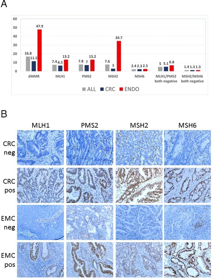
Figure 1.
(A) Distribution of dMMR proteins in the entire cohort and per tumour type. (B) Representative examples of MMR immunohistochemistry in CRC and EMC. Indicated are negative and positive tumours in rows for each MMR protein in columns. In negative tumours, note positive stromal cells and lymphocytic infiltrates (positive endogenous control). For all pictures, original magnification ×200. CRC, colorectal carcinoma; dMMR, mismatch repair deficient; EMC, endometrial carcinoma.

