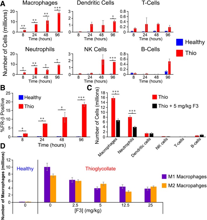
Fig. 3.
Effect of anti-mouse FR-β mAb (F3) treatment on peritoneal immune cell populations following induction of peritonitis with thioglycollate. a Mice were injected intraperitoneally with thioglycollate (Thio), and the immune cell populations were quantitated at the indicated times by flow cytometry. b The percentage of peritoneal macrophages that are FR-β positive is shown as a function of time in both healthy and thioglycollate-treated mice. c Thioglycollate was administered to mice as in panel a, and F3 (5 mg/kg) was injected intraperitoneally 48 h later. After an additional 48 h, the peritoneal cavity was washed and different immune cell populations were quantitated via flow cytometry. d Mice were treated and peritoneal fluid was examined as in panel c except various concentrations of F3 was administered. For all graphs, error bars represent SEM and *, **, and *** denote p values < 0.05, < 0.005, 0.0005, respectively for all panels

