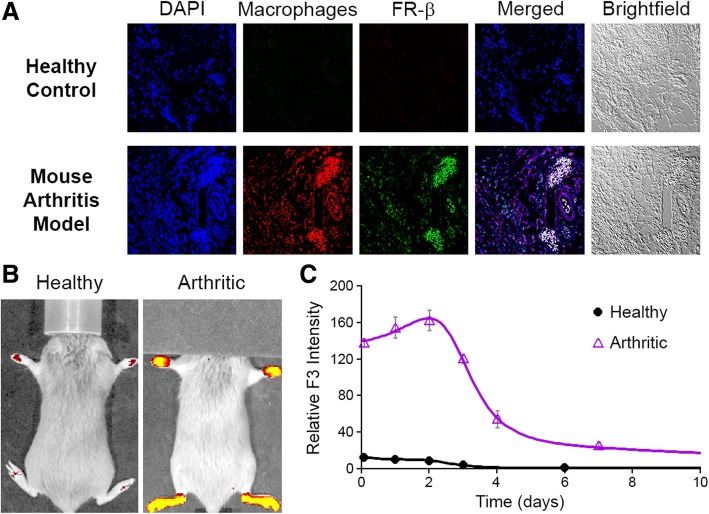
Fig. 4.
Characterization of F3 binding to FRβ-expressing macrophages in arthritic joints. a Two weeks after initial appearance of symptoms in collagen-induced arthritic mice, arthritic joints were sectioned, stained with DAPI dye (nucleus, blue), AF594 dye conjugated anti-F4/80 antibody (macrophage marker, red), and APC dye conjugated anti-FR-β antibody F3 (FR-β, false colored green), and then imaged via fluorescent microscopy. b Mice with collagen-induced arthritis were injected intraperitoneally with AF647-conjugated F3 (50 mg/kg) when the average arthritis score of all 4 paws reached ~ 2. Whole animal fluorescence was imaged at various times post-F3 injection (representative image at 2 days post F3 administration) and paw fluorescence was quantitated as indicated (c)

