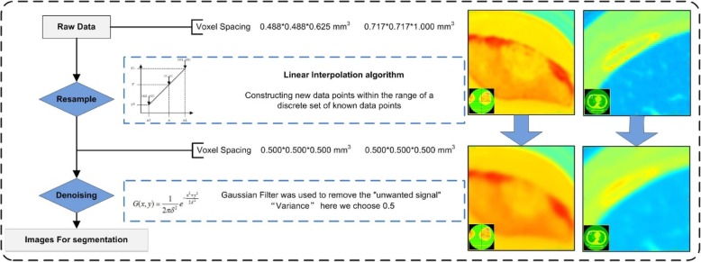
Fig. 2.
Flowchart for data preprocessing. We selected a left knee CT and a chest CT, and correspondingly the sites of tumor segmentation were located in the patella and rib, respectively. The voxel spacing of left knee CT and chest CT were 0.488*0.488*0.625 mm3 and 0.717*0.717*1.000 mm3. By resampling, their voxel spacing were both 0.500*0.500*0.500 mm3. Then denoising was used to get the images for segmentation

