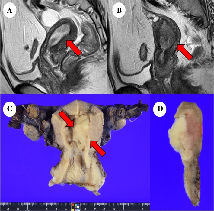
Fig. 1.
Magnetic resonance imaging and macroscopic analysis: (a) T2-weighted image of the initially diagnosed tumor (red arrow). (b) T2-weighted image of the recurred tumor (red arrow). (c) The endometrial mass (red arrows) was 40 × 23 mm-sized in the left wall of the uterine body and (d) had yellow-whitish cut surface

