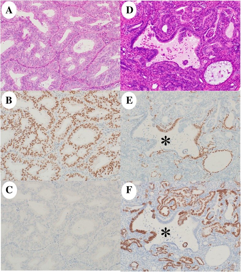
Fig. 3.

Histology and immunohistochemistry of the initially diagnosed tumor. The tumor had (a, 20×) a low-grade endometrioid carcinoma component and (d, 20×) a mesonephric-like adenocarcinoma component. Immunohistochemically, the endometrioid carcinoma was diffuse positive for (b, 20×) estrogen receptor (ER) and negative for (c, 20×) thyroid transcription factor 1 (TTF-1). The mesonephric-like adenocarcinoma showed a transition pattern with a mixture of cells positive for (e, 20×) ER- (a Müllerian duct marker) and (f, 20×) TTF-1- (a Wolffian duct marker) within the same glands (asterisk)
