Fig. 3.
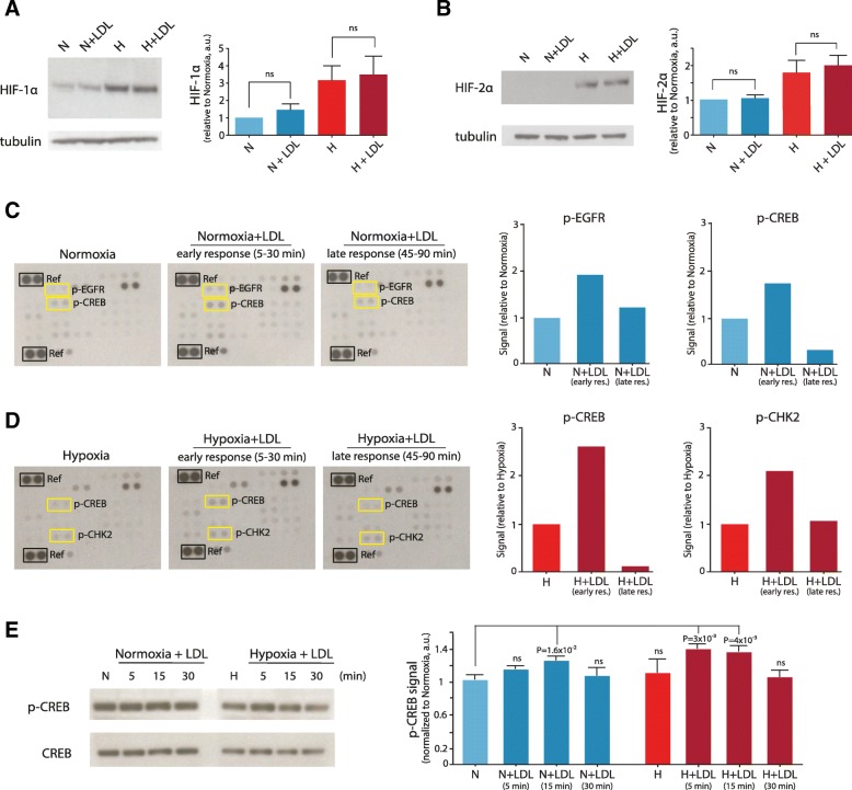
Effects of lipid loading on HIFs and kinase phosphorylation. Western blot analysis of HIF-1α (a) and HIF-2 α (b) expression in lysates of GBM cells (U-87 MG) grown at normoxic (N) or hypoxic (H) conditions for 24 h in the absence or presence of extracellular lipid (+LDL). Shown are representative blots (left panels) and quantifications (right panels) from three independent experiments. Data are presented as the mean fold of normoxic cells ± SD. Phosphokinase antibody array analysis of lysates from normoxic (c) and hypoxic (d) GBM cells following no treatment or treatment with extracellular lipid (+LDL) short-term (5, 15, and 30 min) or long-term (45, 60, and 90 min). Shown are representative blots (left panels) and quantifications (right panels) for p-EGFR, p-CREB and p-CHK2. e, Western blot p-CREB and total CREB analysis of lysates from normoxic and hypoxic GBM cells following no treatment or treatment with extracellular lipid (+LDL) for the indicated time periods. Shown is a representative blot (left panel) and quantification (right panel) from three independent experiments. Quantification of the protein bands was performed by densitometry using ImageJ, and data were expressed as the ratio between p-CREB to total CREB, and then normalized to normoxic conditions without LDL (CTRL), and are presented as the mean fold hypoxia vs normoxia ± SD

