Fig. 5.
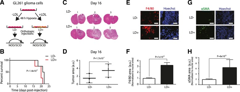
Lipid loading in hypoxic tumor cells promotes angiogenesis and macrophage infiltration in glioma. a, GL261 mouse GBM cells were pre-incubated in hypoxia without (LD−) or with extracellular lipid (LD+) for 48 h, and then injected (50.000 cells/animal) into the brains of NOD/SCID mice. b, Kaplan-Meier plot shows survival in LD− and LD+ groups (N = 5 per group; log-rank test, P = 0.141). c, H&E staining of brains from the LD+ and LD− groups at day 16 post-injection and quantification (d) of the largest tumor area (shown are sections from 3 representative animals from each group). e, Immunofluorescence staining for the macrophage marker F4/80 (red) and Hoechst nuclei (blue) on frozen tumor sections at day 16, and quantification (f) relative to nuclei area (from 3 representative animals per group, 18 fields per animal). Scale bar, 150 μm. g, αSMA (green) and Hoechst nuclei (blue) staining at day 16 and quantification (h) relative to nuclei area (from 3 representative animals per group, 9 fields per animal). Scale bar, 150 μm

