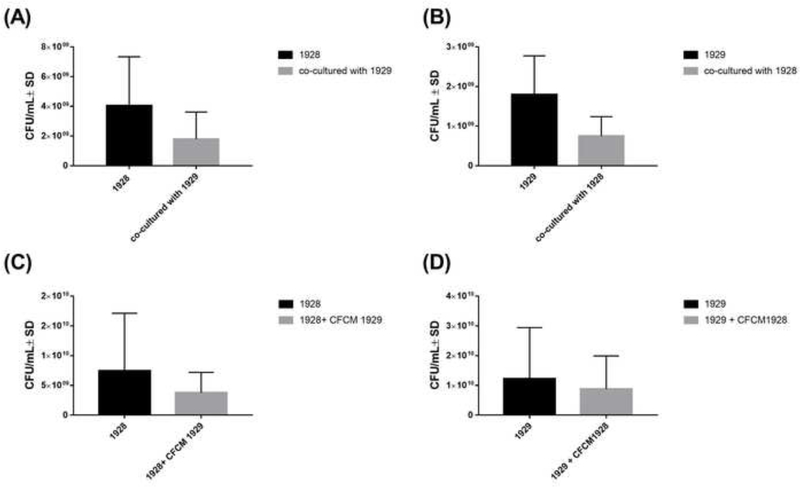
Figure 1. S. aureus 1928 and A. baumannii 1929 growth patterns.
(A) CFU/ml of S. aureus 1928 mono and co-culture with A. baumannii 1929. (B) CFU/ml of A. baumannii 1929 mono and co-culture with S. aureus 1928. (C) CFU/ml of S. aureus 1928 with or without A. baumannii 1929 CFCM. (D) CFU/ml of A. baumannii 1929 with or without S. aureus 1928 CFCM. Bars and whiskers represent means ± SD of three independent experiments in duplicate. The differences between all groups of isolates were analyzed by unpaired t test.

