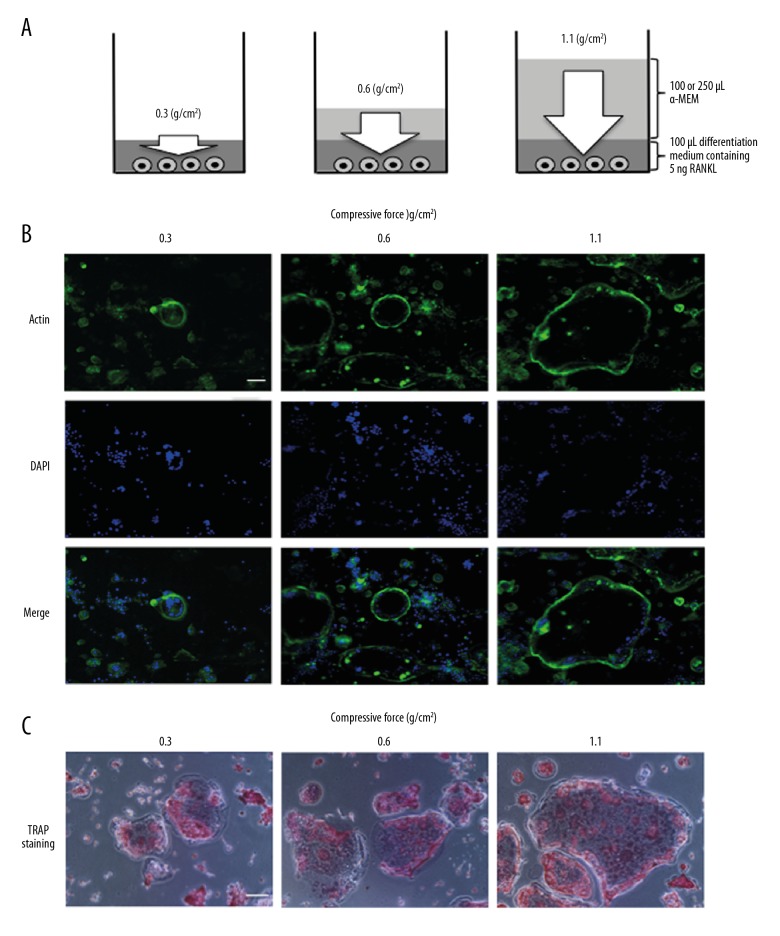
Figure 1.
Schema of compressive force generated by increasing the volume of culture medium and effect of compressive force on actin ring organization and TRAP staining. White arrow indicates compressive force (A). Cells were continuously stimulated with 0.3, 0.6, or 1.1 g/cm2 compressive force in the presence of 5 ng of RANKL for 4 days. Actin labeled with fluorescently tagged phalloidin (green) and nuclei labeled with DAPI (blue) were observed with a fluorescence microscope (B). The cells were stimulated with 0.3, 0.6, or 1.1 g/cm2 compressive force for 4 days, and then stained with TRAP and observed by light microscopy (C). Scale bar=100 μm.

