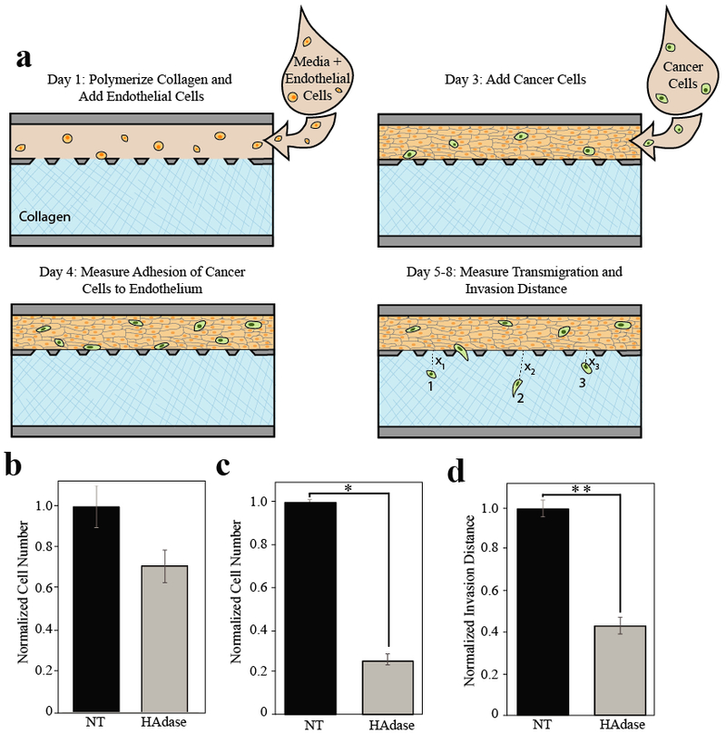
Figure 3. Removal of pericellular HA reduces tumor cell metastatic potential in vitro.

a) Timeline of experimental process. On day one unpolymerized collagen solution (blue) was perfused into larger of two adjacent channels. After collagen was polymerized, media and endothelial cells (tan) were added to the perfusion channel. Devices were then placed in a 37°C incubator for 24-48 hours until endothelial cells formed a confluent monolayer. On day three, cancer cells (green) were added to the perfusion channel under constant flow. Media was perfused for a total of 5 days after cancer cells were added, and each device was imaged every 24 hours. Adhesion was quantified by counting cells adhered in the endothelial channel 24 hours after cancer cells were added. The number of cells that transmigrated and the average distance of migration was measured (ie. Average distance = (Σ X1+ X2+ X3+… Xn)/n) using ImageJ to obtain the distance from the edge of the endothelium to the middle of the cell body. b-d) Quantification of metastatic potential of MDA-MB-231 breast carcinoma cells with and without hyaluronidase treatment. c) Adhesion of cancer epithelial cells to endothelium. There is no significant difference in endothelial adhesion between hyaluronidase (HAdase) treatedand untreated (NT) tumor cells. d) Transmigration between treatment groups was statistically significant, p < 0.0005. e) The average invasion distance also showed a statistiaclly significant difference across treatment groups (*p < 0.0005 and **p < 0.007). All experiments were performed at least three separate times with at least n=3 in each treatment group. Data was then normalized within each experiment and experiments were averaged before comparison.
