
Extended Data Fig. 2 |. Expression and plasma-membrane localization of PCDH1 variants lacking domains EC1 or EC2 of PCDH1.
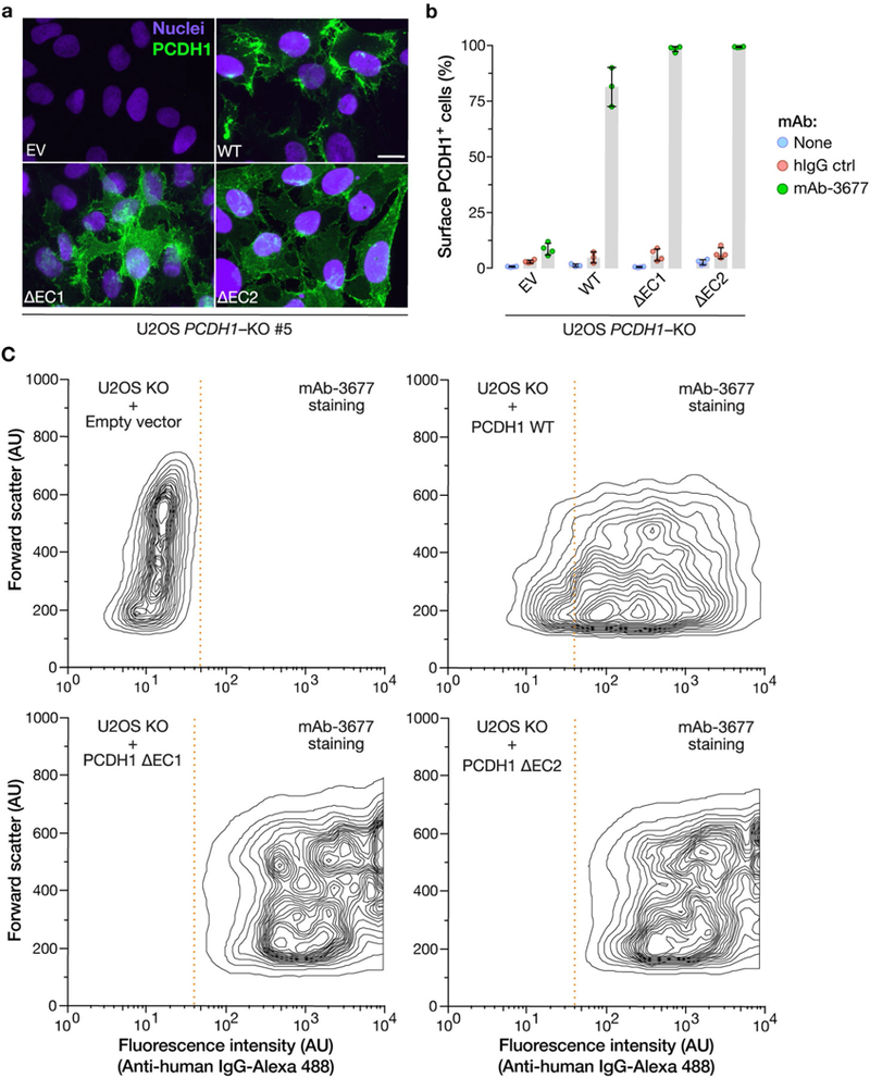
a, U2OS PCDH1-KO cell lines complemented with the indicated PCDH1 proteins were immunostained with an anti-Flag antibody and visualized by fluorescence microscopy. EV, empty vector. Scale bar, 20 μm. b, c, Live cells from a were stained with the PCDHl-EC7-specific mAb 3677 at 4 °C to detect cell-surface PCDH1 and visualized by flow cytometry. Cells were gated on PCDH1 immunofluorescence intensity (dotted red lines in c) to determine the percentage of cells with surface expression of each PCDH1 protein. Averages ± s.d. are shown in b: two experiments, n = 4, except in the case of WT, for which n = 3. c, Representative flow plots from b. Experiments were performed three times with similar results.