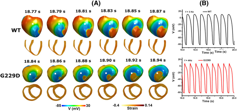
FIGURE 11.
Reentrant dynamics wave in the 3D ventricular tissue model for the wild type and G229D mutation conditions. A, Snapshot of the transmural distribution of membrane potential (top) and strain (bottom). B, Time traces of action potentials at the center of the ventricle (white dot). (top) WT condition and (bottom) G229D condition

