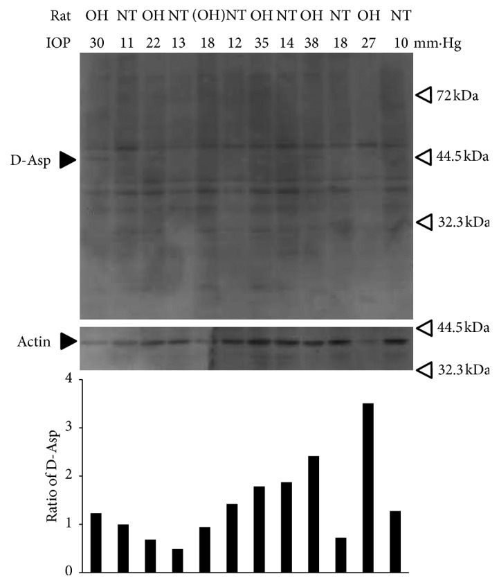
Figure 2.

Replicability of the induction of proteins including D-aspartic acid in the retina of ocular hypertensive and normotensive rats. Total lysates of retinas derived from 6 rats with OH or NT eyes blotted with anti-D-aspartic acid antibody and anti-actin antibody. Black arrow, labelled D-Asp, shows the protein band including D-aspartic acids. The samples of the first and second lanes are the same as in Figure 1(a) and are blotted as a positive control. The fifth and sixth lanes were derived from a rat which failed to achieve glaucoma status (defined as IOP > 21) and was blotted as a negative control. Lowest panel, which shows the ratios of the expression volumes of proteins containing D-aspartic acid corrected with each actin expression, showed that protein bands in the first, third, ninth, and eleventh lane had a stronger protein band including D-aspartic acid around 44.5 kDa than control NT.
