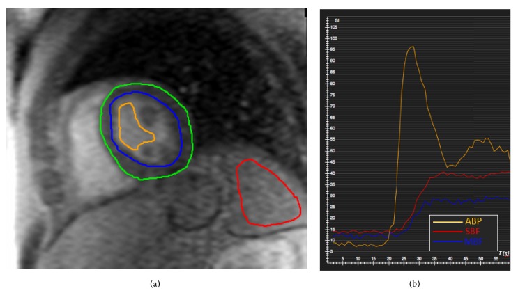
Figure 2.
(a) Signal intensity (SI) values were measured in each time frame of the perfusion sequence by drawing 9 cm2 regions of interest (ROI) within the spleen parenchyma (red line) to calculate SBF. ROIs within the left ventricle cavity (orange line) were drawn for ABP quantification. To assess MBF, SI of the whole myocardium between epicardial (green) and endocardial (blue) layers was measured. (b) The graph shows the SI-to-Time curves of ABP (orange), SBF (red), and MBF (blue). ABP= arterial blood pool; SBF= splenic blood flow; MBF= myocardial blood flow.

