Figure 7.
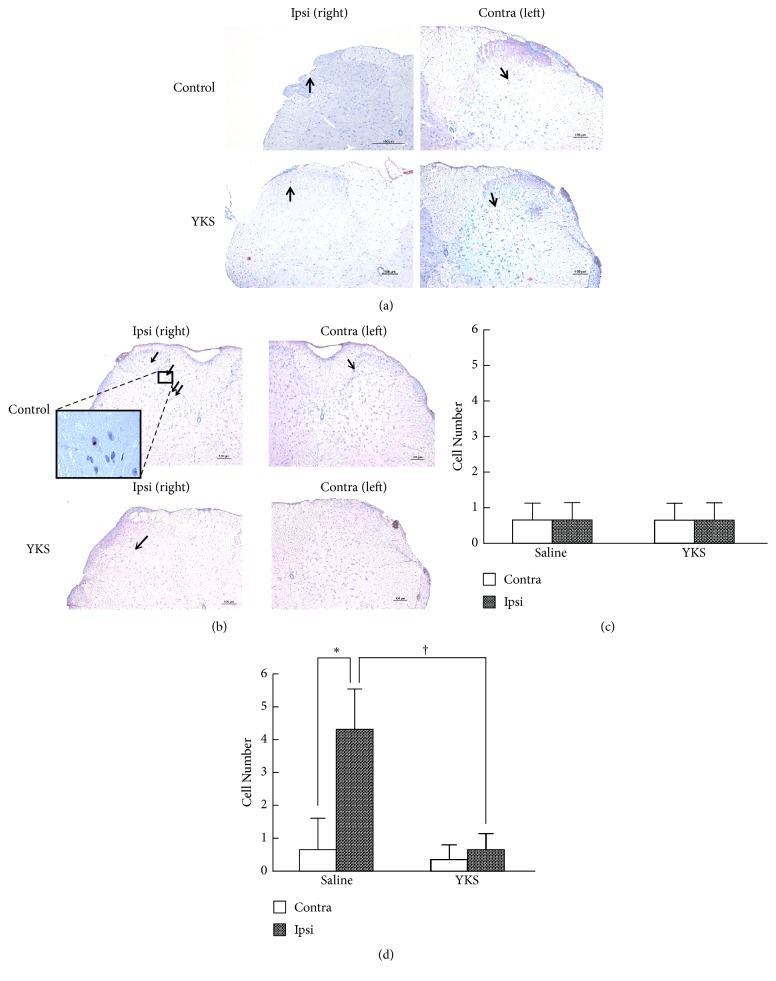
MMP-9 and MMP-2 immunostaining in the spinal cord. (a, b) Immunostaining of MMP-2 (a) and MMP-9 (b) in the dorsal horn of the spinal cord. The lumbar spinal cord (L4-L5) was prepared from cancer pain model mice on day 7 following p.o. administration of saline or YKS (10 mg) once daily. Transverse sections (40-μm thick) were immunostained with anti-MMP-2 and MMP-9 antibodies as described in “Materials and methods.” The sections were counterstained with hematoxylin. Scale bars, 100 μm (a, b). Arrows indicate immunopositive cells. (c, d) The number of MMP-2- (c) and MMP-9- (d) positive cells in the dorsal horn. The number of immunopositive cells was counted in 4 sections for each treatment. The data are expressed as the mean ± SD (n = 4). Student's t-test was applied for statistical analysis. ∗P<0.05, compared to left part. †P < 0.05 compared to YKS.

