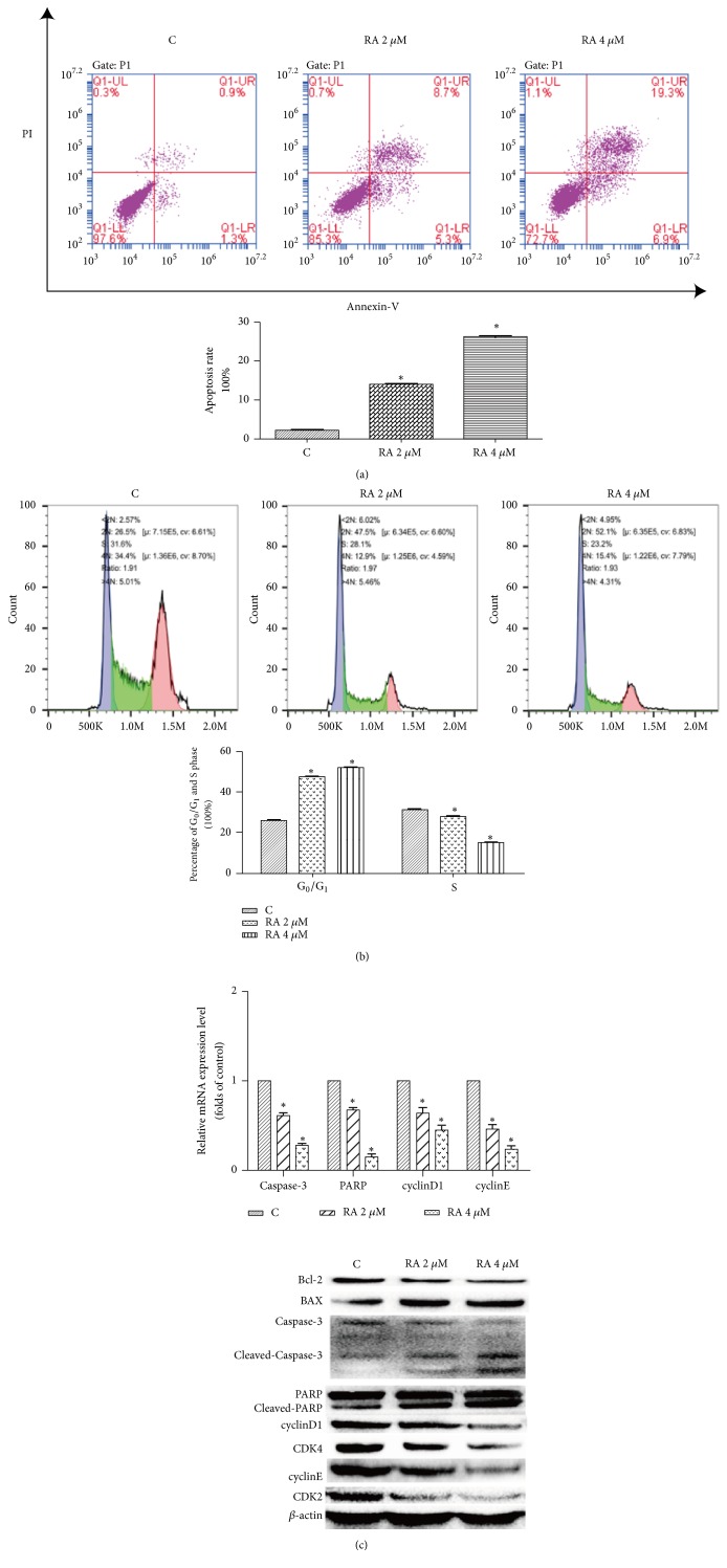
Figure 2.
Raddeanin A (RA) induced apoptosis and blocked cell cycle progression in HCT116 cells. (a) After treatment with RA (2 and 4 µM) for 12 h, the cells were incubated with annexin-V-FITC and propidium iodide (PI) and the apoptosis rate was analyzed via flow cytometry. The results shown are from a representative experiment. (b) Representative results of cell cycle analysis conducted by flow cytometry in HCT116 cell treated with different doses of RA for 12 h (n = 3). (c) The expression of apoptotic proteins and cell cycle-related factors was detected by RT-PCR and western blotting. The data shown are the mean ± SD (n=3, ∗P<0.05, compared with the control). β-actin was used as an internal control.

