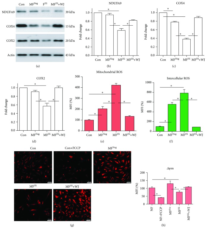
Figure 4.
Mitochondrial malfunction of MELAS fibroblasts is improved following mitochondrial transfer. (a) Representative immunoblot image of mitochondrial OXPHOS subunits (nuclear-encoded NDUFA9 and COX4 and mtDNA-encoded COX2). Actin as loading control. (b–d) Quantitative results of protein expression level normalized to actin. (e, f) Mitochondrial and intracellular ROS detected with MitoSOX™ Red and H2DCFDA, respectively. (g) Mitochondrial membrane potential (ΔΨm) measured by TMRE stain and photographed using a fluorescence microscope. FCCP used to dissipate ΔΨm served as negative control. Scale bar, 100 μm. (h) Mitochondrial membrane potential quantitatively analyzed using flow cytometry. ∗ p < 0.05 significantly different when compared to the indicated group. WJ: Wharton's jelly mesenchymal stem cell; Con: control fibroblast from normal human; MFNeg: MELAS fibroblast clone harboring negative mutation burden; MFHi: MELAS fibroblast clone harboring high mutation burden; MFI: mean fluorescence intensity.

