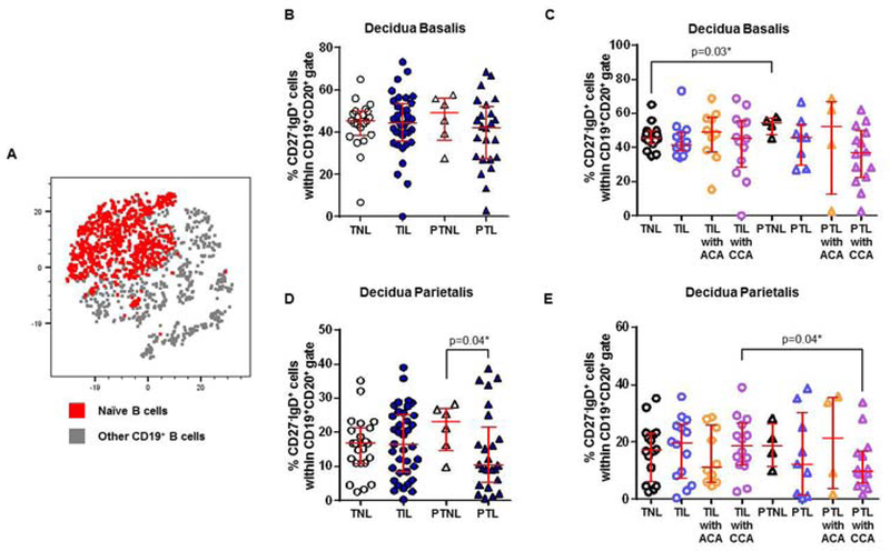
Figure 5. Naïve B cells in the decidua basalis and decidua parietalis.

(A) A representative t-distributed stochastic neighbor embedding (t-SNE) dot plot visualizing naïve B cells in the decidual tissues. Red = naïve B cells and grey = other CD19+ B cells. The proportions of naïve B cells in the decidua basalis (B) or decidua parietalis (D) from women who delivered at term with labor (TIL) or without labor (TNL) and women who delivered preterm with labor (PTL) or without labor (PTNL). N = 6 – 37 per group. The TIL and PTL patients were subdivided into those with acute histologic chorioamnionitis (ACA) or chronic histologic chorioamnionitis (CCA), and those without these lesions. Non-labor controls without ACA or CCA were included as well. The proportions of naïve B cells in the decidua basalis (C) or decidua parietalis (E) in these patient subgroups. N = 4 – 16 per group. Red midlines and whiskers indicate medians and interquartile ranges, respectively.
