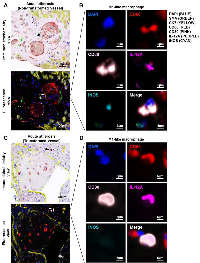
Figure 5. Immunolocalization of M1-like macrophages in the decidual vessels with acute atherosis.
Multiplex immunofluorescence staining showing nuclear staining (4′,6-diamidino-2-phenylindole, DAPI, blue), smooth muscle actin (SMA, green), cytokeratin-7 (CK7, yellow), CD68 (red), CD80 (pink), interleukin (IL)-12A (purple), and inducible nitric oxide synthase (iNOS, cyan) in the decidua basalis with acute atherosis. Representative images showing (A) the immunohistochemistry and fluorescence views and (B) magnification of an M1-like macrophage in the vessel wall of a non-transformed decidual vessel with acute atherosis. Representative images showing (C) the immunohistochemistry and fluorescence views and (D) magnification of an M1-like macrophage near a transformed decidual vessel with acute atherosis. Phenoptics was performed to generate separate and merged immunofluorescence images (B&D), and to convert fluorescence images to the immunohistochemistry view (A&C). Black arrows in the immunohistochemistry view and dotted boxes in the fluorescence view indicate an M1-like macrophage. Images are representative of 3 experiments per group. Images were taken at 200X magnification, and a close-up of an M1-like macrophage is shown. Scale bars = 50μm (original image) or 5μm (close-up image).

