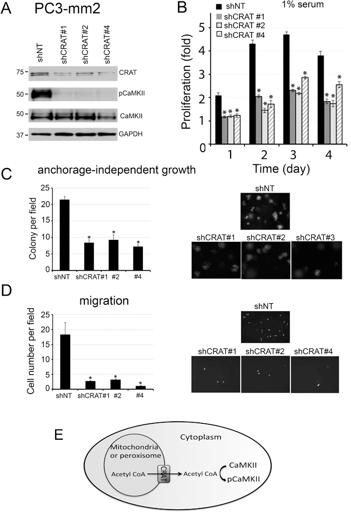
Figure 5. Knockdown of CRAT in PC3-mm2 cells.

CRAT in PC3-mm2 cells was knocked down by using shRNAs #1, #2 or #4 in lentiviral vectors. (A) Total cell lysates were immunoblotted for CRAT, pCaMKII, and total CaMKII. GAPDH was used as a loading control. (B) Cells were cultured in 1% FBS. Cell proliferation is expressed as fold change of cell number compared to day 0. n=3. The doubling time of shNT was 23.0 ± 1.1 hours and the shCRAT #1, #2 and #4 were 29.5 ± 1.6 hours, 41.4 ± 2.3 hours and 32.9 ± 1.1 hours, respectively. The plating efficiency, determined at 24 hours after cell seeding, of shNT, shCRAT #1, #2 and #4 were 210.0%, 117.3%, 120.0% and 123.0% respectively. (C) Cells were grown in soft agar. Number of colonies per field was counted. n=3. (D) Cells were seeded onto a Boyden Chamber. Cells migrated through the membrane were labeled with Calcein AM. n=2. (E) Schematic illustration for a role of CRAT, expressed in peroxisome or mitochondria, in the transport of acetyl-CoA to the cytosol for CaMKII activation. *, p<0.05.
