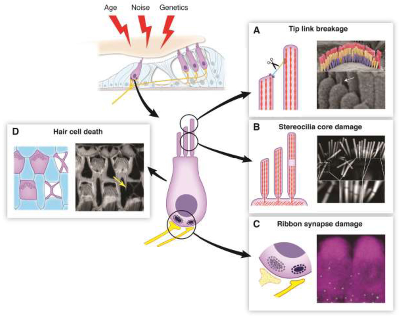
Figure 1, Key Figure. An Overview of Hair Cell Damage.
(Top Left) Hair cells can accumulate damage stemming from a variety of factors including age, noise, and genetics. (Center) Diagram of a hair cell indicating sites vulnerable to damage. (A) Tip link breakage. Left: Tip links can be broken by overstimulation or by in vitro calcium chelation, leading to a loss of tension on the MET channel complex and subsequent loss of the MET current. Right: Scanning electron micrograph showing an outer hair cell bundle. Tip links (white arrow) are visible at higher magnification below. (B) Stereocilia core damage. Left: Overstimulation of hair bundles causes the appearance of gaps in staining of the stereocilia F-actin core. These gaps likely represent sites of F-actin depolymerization, which would decrease bundle rigidity. Right: Phalloidin staining of a noise-damaged inner hair cell bundle. Gaps in the staining are visible at higher magnification below. (C) Ribbon synapse damage. Left: Ribbon synapses can be lost due to exposure to loud noise or prolonged exposure to milder noise, even in absence of permanent hearing threshold shift. Their loss can reduce hearing ability in noisy environments, known as “hidden hearing loss”. Right: Immunolabeling of CTBP2 (a component of ribbon synapses, green puncta) at the base of hair cells labeled by MYO7A immunostaining (magenta). (D) Hair cell death. Left: Auditory sensory epithelium with F-actin scars at the sites of missing hair cells. When hair cells die, they are extruded from the epithelium. Nearby supporting cells fill in the hole, leaving a cross-shaped F-actin scar. Right: Phalloidin labeling of F-actin in the organ of Corti from an aged mouse. An F-actin scar is seen at the site of a missing hair cell (yellow arrow). All images were taken in the Shin lab.

