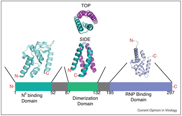
Figure 2:
Schematic representation of the modular organization of the RABV phosphoprotein (P). The N0 binding domain is teal, the dimerization domain is green, and the ribonucleoprotein binding domain (RNP) is periwinkle. The solved crystal structure for the N0 binding domain is depicted in teal (PDB 3OA1). The solved crystal structure for the dimerization domain is depicted in green and pink with both top and side views (PDB 3L32). The solved structure for the RNP is depicted in periwinkle (PDB 1VYI). [19, 127, 135, 137, 138]

