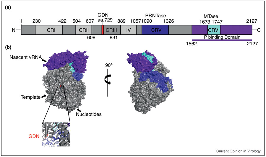
Figure 3:
A) Schematic diagram depicting the domain organization of RABV large protein (L). The GDN polymerization motif is in red. The polyribonucleotidyltransferase (PRNTase) is in blue. The methyltransferase (MTase) is in cyan. The phosphoprotein (P) binding region is purple. Conserved regions (CR) of the non-segmented negative-sense RNA viruses are labelled CR I -VI. B) Surface representation of the RABV L generated by homology modelling based on the coordinates reported for the closely related VSV L structure with the same color scheme as described by 3A. Below is a zoomed in ribbon representation of the GDN motif responsible for polymerase activity. [25, 33, 36, 128]

